Author name: Dr Bradley

Dr. Timothy Bradley, a distinguished member of the Pediatric Orthopedic Society of North America and the American Academy of Orthopedic Surgeons, is a leading expert in pediatric orthopedics serving the community of Brandon, Riverview and whole of Tampa. With a commitment to providing the highest quality care for young patients, Dr. Bradley has earned a reputation for excellence in the field. His expertise and dedication to improving the lives of children through orthopedic solutions make him a trusted authority in the orthopedic community. Dr. Bradley's  work at POBAR in Tampa exemplifies his passion for compassionate and effective healthcare, ensuring a brighter and healthier future for young patients.

A POBAR pediatric orthopedic specialist gently guiding a child with joint concerns through safe activity, focusing on Osteochondritis Dissecans care in Brandon.
Joint Concerns

Helping Your Child Stay Active Despite Joint Concerns

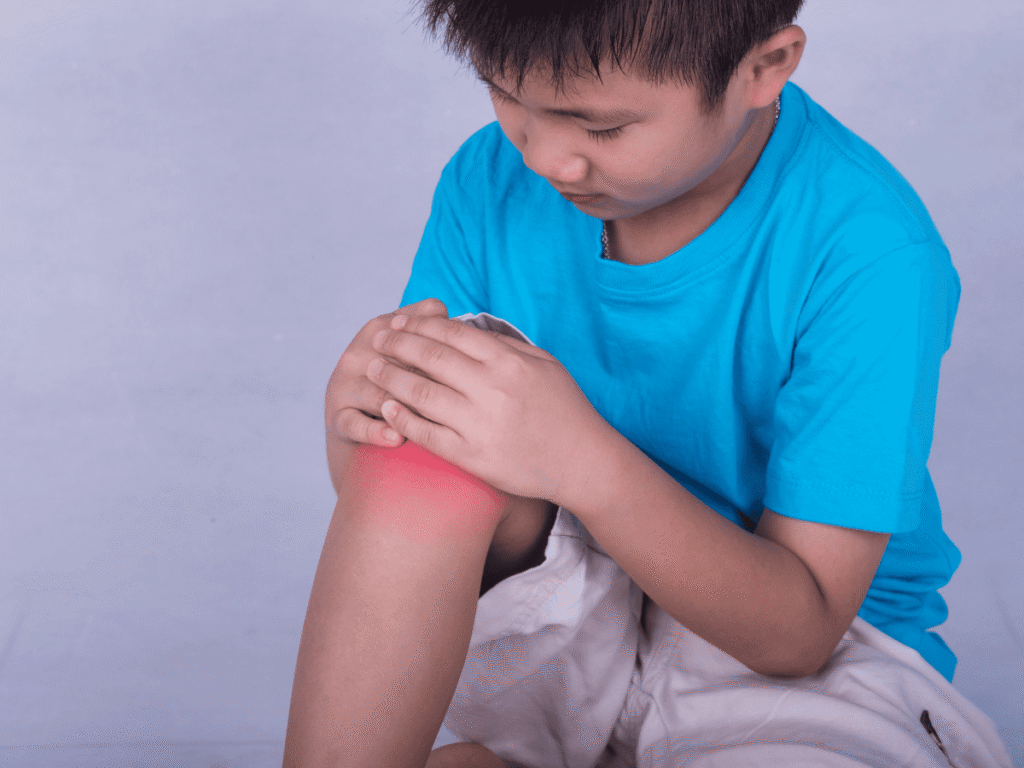
Helping your child stay active despite joint concerns like Osteochondritis Dissecans (OCD) begins with understanding the condition and giving your family the tools to navigate it with confidence. Children and teens involved in sports may develop this condition when a piece of cartilage and underlying bone separates from a joint surface, most often in the knee but also in the elbow or ankle. This can cause pain, swelling, and difficulty with movement that may impact participation in play and athletic activities. If you notice persistent joint pain, swelling, or changes in how your child moves, reach out to POBAR to learn how we can help your child stay engaged in life and play. Children who love sports and play may show subtle changes before more obvious symptoms of Osteochondritis Dissecans appear, and those early signs are often the key to preserving joint health while keeping your child active. The discomfort they experience during running, jumping, or pivoting might lead them to stop participating or limp when they walk — signals that merit attention from a pediatric orthopedic specialist trained in OCD care. Rather than simply signaling the end of activity, these early changes offer an opportunity to adjust your child’s routine in ways that protect their joints and still allow for fun and growth. With the right plan, most children can continue to enjoy age‑appropriate movement, even if that looks different from their initial sports schedule. Understanding Osteochondritis Dissecans in Children Osteochondritis Dissecans in children is a joint condition where a fragment of bone with its overlying cartilage becomes weakened and may detach due to interrupted blood supply and repetitive stress on a joint during growth. The knee is the most commonly affected joint, but the elbow and ankle are also susceptible, particularly in young athletes who engage in repetitive motions and high‑impact activities. Symptoms often include pain during activity, swelling around the joint, and sometimes a sense that the joint is catching or locking, which can understandably lead to frustration in an active child. The severity of symptoms and how they affect your child’s daily activities depends on how large the lesion is and whether the affected piece of bone and cartilage remains stable. Diagnosis typically involves a pediatric orthopedic evaluation that includes a physical exam and imaging studies such as X‑rays, which help visualize the bone and joint. In many cases, an MRI is also used to assess how the cartilage and subchondral bone are involved and whether any loose pieces are present within the joint space. These studies give specialists a clear picture of your child’s joint health and help guide individualized treatment plans. What Causes Osteochondritis Dissecans? While the precise cause of OCD remains partly unknown, most experts agree it is linked to repetitive stress or microtrauma to the joint during periods of rapid growth. Young athletes who participate in activities that involve frequent running, jumping, or throwing may be particularly at risk. There may also be a genetic component or variations in joint development that influence how susceptible a child is to this condition. Additionally, periods of rapid skeletal growth during childhood and adolescence may make certain joints more vulnerable to disruptions in blood flow that predispose to an OCD lesion. Although these risk factors can increase the likelihood of an OCD lesion, many children with joint symptoms experience only mild changes that respond well to careful activity modification and monitoring rather than immediate surgery. Understanding these factors can empower parents to make thoughtful decisions about activity levels and rest. Recognizing Symptoms That May Affect Activity Children with Osteochondritis Dissecans may exhibit several symptoms that can interfere with normal play and sports participation: Pain with Activity: The most common sign is discomfort that increases with weight‑bearing activity and may improve with rest. Swelling and Tenderness: Some children experience noticeable swelling around the affected joint after exercise or prolonged use. Decreased Range of Motion: Your child might have difficulty fully bending or straightening the knee, elbow, or ankle. Catching or Locking: Sensations of the joint locking or catching during movement suggest that a fragment may be interfering with smooth motion. Instability: A feeling that the joint is “giving way” can make your child hesitant to participate in activities that require sudden changes in direction. These symptoms often prompt families to reduce activity to avoid pain. However, the goal is to find a balance that protects the joint without completely isolating your child from movement, which is essential for overall health and wellbeing. How to Help Your Child Stay Active Safely Supporting your child’s desire to stay active while managing Osteochondritis Dissecans involves strategic adjustments that protect the affected joint and promote healing: Activity Modification: Instead of high‑impact sports, encourage low‑impact activities like swimming or cycling, which strengthen muscles without excessive stress on the joints. Proper Warm‑Up and Cool‑Down: Ensuring your child warms up before play and cools down afterward can reduce joint stress and help maintain flexibility. Strength and Flexibility Exercises: Physical therapy focused on strengthening the muscles around the joint can improve stability and support healthy movement. Structured Rest: Incorporating rest periods into your child’s routine helps reduce inflammation and gives injured cartilage and bone a chance to recover. Protective Gear: Using braces or supportive padding during high‑risk activities may provide additional joint support without completely stopping activity. These approaches emphasize keeping your child engaged in movement that feels good and is appropriate for their stage of healing, rather than eliminating activity entirely. Your pediatric orthopedic specialist can help you tailor these strategies based on your child’s symptoms and lifestyle. When to Consider Specialist Intervention If your child’s symptoms persist despite activity modification and supportive care, or if imaging studies show a loose fragment of bone within the joint, our pediatric orthopedic specialist may recommend more structured interventions. In growing children, many OCD lesions can heal with conservative approaches like rest, temporary immobilization, and gradual return to activity under guidance. However, if the lesion doesn’t heal or pain continues, surgical options such as drilling

Gait Abnormalities

Early Signs of Arm or Leg Differences and What to Do Next

Children’s arms or legs that look different from what you expect can be unsettling for any parent, but recognizing early signs of arm or leg differences can make a profound difference in your child’s health and development. Visible curvature, limb-length differences, or unusual gait patterns may be early indicators of underlying orthopedic issues that benefit from expert evaluation. At POBAR, we offer gentle, child‑friendly assessment and personalized care to guide families through diagnosis and treatment options with compassion and clarity. If you notice any signs that concern you, reach out to our pediatric orthopedic team to learn how we can help your child thrive. From the moment you notice one limb looking different from the other, your caregiving instincts kick in—so you’re not alone in that concern about early signs of arm or leg differences. Some children may have visible asymmetry, bowed limbs, or an irregular way of walking that seems “off,” and these signs can point to conditions ranging from growth variations to congenital differences. Understanding what to watch for and what steps to take next empowers you to pursue the right care early. Being observant now sets the stage for confident decision‑making about your child’s musculoskeletal health. What Are Early Signs to Watch For? When parents notice something unusual about a child’s arms or legs, it can trigger worry—but understanding which signs suggest the need for specialist evaluation helps you act with reassurance. Early signs of arm or leg differences often involve visible asymmetry, such as one arm or leg appearing shorter or more bowed than the other, uneven shoulder or hip height, or an unusual gait when your child walks or runs. These findings may be subtle at first, but if they persist or are associated with discomfort, reduced mobility, or changes in how your child uses the limb, a pediatric orthopedic assessment is recommended. Some children may also show restricted range of motion or have difficulty reaching, climbing, or participating in age‑appropriate activities compared with their peers. These observable patterns are often the first clues that specialist evaluation and imaging studies—like X‑rays or CT scans—can help clarify. Early recognition allows for thoughtful monitoring or intervention before more significant functional concerns develop. Common Patterns That May Indicate an Underlying Issue Not all differences in limb appearance signal a serious problem, but some patterns are worth paying attention to: Limb-Length Discrepancy: One arm or leg noticeably longer or shorter than the other, which can affect posture or gait. Bowed or Knocked Knees: Legs that curve outward or inward beyond typical developmental variation. Angular Deformities: Visible bends or angulation in bones that don’t align with normal growth patterns. Uneven Gait or Movement: Limping, toe walking, or awkward stepping that persists beyond expected developmental stages. Asymmetrical Appearance: Shoulders, hips, or wrists that appear uneven or a limb that “looks different,” regardless of discomfort. While many of these findings are part of normal variation in early childhood, persistent or progressive differences are worth evaluation. Pediatric orthopedic specialists are trained to distinguish between typical developmental patterns and those that may require monitoring or intervention. Why Early Recognition Matters Spotting early signs of arm or leg differences isn’t just about appearance—it’s about your child’s overall function and comfort. Left unaddressed, some limb differences can lead to pain, compensatory movement patterns, or limitations in sports and everyday activities. For example, a significant limb-length discrepancy can alter how your child walks and may contribute to joint discomfort if left untreated. Likewise, angular deformities like bowed legs that don’t resolve with age may point to an underlying growth issue, such as rickets or growth plate disturbance, which benefits from early specialist care. Recognizing these patterns early allows healthcare professionals to tailor a plan that may include observation, bracing, guided growth techniques, or surgery, depending on your child’s unique needs. What to Do Next: Path to Evaluation and Diagnosis If you see signs that concern you, the next step is compassionate, comprehensive assessment by a pediatric orthopedic team. Our process begins with a detailed physical examination and a thoughtful conversation about your child’s development and symptoms. Advanced imaging, such as X‑rays or CT scans, may be used to understand the shape and alignment of bones in the arms or legs. These studies offer a clearer picture of how the bone is growing and whether there is a structural difference. After diagnosis, a personalized treatment plan is developed with your family’s goals and your child’s functional needs at the center. Understanding Possible Causes Early signs of arm or leg differences can arise from a variety of underlying causes, and understanding these helps set expectations for care. Some children may have congenital differences present at birth due to conditions that affect limb formation, like fibular hemimelia or radioulnar synostosis. Other differences occur from growth disturbances or injuries to growth plates, which can alter how bones lengthen over time. Conditions such as rickets, caused by vitamin D deficiency, can lead to bowing of the legs, while genetic or skeletal dysplasia disorders may result in shortened or curved limbs. Every child’s story is unique, and a careful diagnostic approach ensures that the right factors are considered. Treatment Options Tailored to Your Child What treatment looks like depends on your child’s age, the cause of the limb difference, and how much it affects function or comfort. In many cases, non‑surgical options are effective and child‑centered: Observation and Monitoring: Some variations resolve naturally as your child grows. Orthotic Support: Braces or splints can gently guide limb alignment during growth phases. Guided Growth Techniques: These are minimally invasive procedures that use small implants to influence how bones grow, helping correct angular differences over time. For more significant or progressive differences, surgical correction may be recommended to realign bones, address length discrepancies, or improve function. Regardless of the path chosen, your child’s care team at POBAR works closely with you to explain each option, recovery expectations, and long‑term growth monitoring to ensure the best outcomes. POBAR Pediatric Orthopedic Care: Supportive, Skilled, and Family‑Focused At

Pediatric orthopedist explaining meniscus tear care to a young patient’s parent at POBAR clinic in Brandon
Meniscus Tear

Knee Knots: What a Meniscus Tear Means for Your Child

A meniscus tear can leave your child’s knee feeling stiff, painful, or unstable, especially after a sudden twist or fall during play. At POBAR, we help families understand what causes a pediatric meniscus tear and guide you through diagnosis, recovery, and prevention with compassionate expertise. Early recognition and thoughtful care can make all the difference in healing and preventing future issues. Contact us to learn how our child‑friendly team supports strong knees and confident kids. From the grassy field of a soccer game to a quick pivot on the playground, children’s knees work hard every day. The meniscus is a C‑shaped piece of cartilage in the knee that acts as a shock absorber and helps distribute weight as your child runs, jumps, and plays. A tear in the meniscus can happen when the knee twists suddenly while bearing weight or during impact—especially in active kids and young athletes. These tears can lead to pain, swelling, stiffness, or a catching sensation in the knee, and understanding what they mean helps you act with confidence.   What Does a Meniscus Tear Really Mean? The meniscus sits between the thighbone and shinbone and cushions the knee joint like a natural shock absorber. When a meniscus tear occurs, that cushioning is disrupted, which can affect your child’s ability to bend, straighten, or bear weight comfortably. Tears range from small frays that lead to mild discomfort to larger or displaced tears that cause catching, locking, or instability in the knee joint. This range of severity means that symptoms can vary quite a bit, and careful evaluation helps ensure your child gets the right care.   Common Causes of a Pediatric Meniscus Tear Meniscus tears in children often happen during activities that involve sharp twists, pivots, or sudden changes in direction—movements common in sports like soccer, football, and basketball. A direct hit to the knee during play or repetitive stress from frequent squatting and kneeling can also contribute to tears. While aging is a common factor for meniscus problems in adults, in children it’s more often linked to active play or collisions, and underlying knee misalignment or instability may make a tear more likely. Common causes may include: Sudden twisting or pivoting of the knee during sports activities Direct impact to the knee from a fall or collision Overuse or repetitive stress on the knee joint Pre‑existing knee alignment issues Participation in high‑impact or contact sports Excessive strain from abrupt movements or uneven surfaces All these movements place stress on the cartilage and can lead to tears if the knee twists beyond its normal range or experiences a forceful hit.   Recognizing the Symptoms in Your Child Understanding how to spot the signs of a meniscus tear can help you seek timely care and support your child’s recovery: Pain: Often felt along the joint line, especially when bending or twisting the knee. Swelling and stiffness: Common soon after the injury, sometimes developing over hours or days. Difficulty bending or straightening the knee: Pain or mechanical blockage may restrict motion. Locking or catching sensation: When a fragment of the torn cartilage gets trapped between joint surfaces. Instability or “giving way”: Especially during activities that require sudden direction changes. These symptoms may vary depending on the size and location of the tear as well as whether other knee structures are also affected.   How a Meniscus Tear Is Diagnosed We begin with a gentle and thorough clinical evaluation to understand your child’s symptoms and knee function. A physical exam guides our assessment, and diagnostic imaging—most commonly an MRI—is often used to visualize soft tissues like cartilage and confirm the extent of the tear. Early and accurate diagnosis ensures that your child receives the most appropriate care for their specific injury.   Treatment Options for Pediatric Meniscus Tears Meniscus tear treatment depends on the severity of the injury, your child’s symptoms, and the tear’s location. Pain and swelling may be managed initially with rest, ice, compression, and elevation, while supportive braces can help protect the knee during early healing. Non‑surgical care may include: Temporary activity modification to reduce stress on the knee Physical therapy to strengthen muscles and improve stability Bracing during activity to support the joint In cases where symptoms persist or the meniscus tear affects knee mechanics significantly, minimally invasive arthroscopic surgery may be recommended. Techniques such as repairing the tear or trimming the damaged cartilage help restore function and promote long‑term knee health. Children often heal well following arthroscopic procedures, especially when paired with tailored rehabilitation.   Helping Your Child Return to Activity After meniscus treatment, whether non‑surgical or surgical, a structured rehabilitation plan helps rebuild strength and flexibility in the knee. Physical therapy focuses on restoring range of motion, strengthening surrounding muscles, and gradually increasing activity levels in a safe, controlled way. Recovery timelines vary with the injury’s severity, but many children are able to return to their favorite activities once strength, stability, and comfort are restored.   Can Meniscus Tears Be Prevented? While no strategy completely eliminates the risk of a meniscus tear, good knee care habits can help reduce the likelihood of injury and support long‑term joint health: Warm up and cool down: Prepares muscles and joints for movement. Use proper footwear: Shoes with good support reduce knee strain. Avoid abrupt twisting motions: Especially on hard or uneven surfaces. Gradual activity progression: Helps muscles adapt to increased demands. Cross‑training: Promotes balanced muscle development. Healthy lifestyle habits: Maintaining a healthy weight supports knee joints. Encouraging safe play and listening to your child’s body—especially if they complain of discomfort during activity—can help protect their knees and keep them active with confidence.   Compassionate Pediatric Knee Care at POBAR At POBAR, we know how much you want your child to stay active, safe, and happy. Our caring team specializes in pediatric meniscus tear evaluation and treatment designed for young athletes and active kids. From thorough assessments to personalized treatment plans and gentle guidance through recovery, we’re here to support your family with expert care and

A caring pediatric orthopedist examining a child with a joint sprain at POBAR clinic in Brandon.
Sprains

Bumps, Twists, and Turns: Understanding Pediatric Sprains

Pediatric joint sprains can be worrying for both children and parents, but with the right care, healing and strength can return. At POBAR, our experts guide families through recognizing symptoms, getting the right diagnosis, and following a treatment plan suited to your child’s needs. Understanding how a sprain happens, what to watch for, and how long recovery takes can ease your mind and help your child feel better sooner. Contact us for personalized guidance, compassionate care, and support at every step. From a playful jump on the playground to spirited games of backyard soccer, childhood is full of bumps and twists. Pediatric joint sprains happen when the strong bands of tissue called ligaments stretch or tear, usually from an unexpected twist, fall, or tumble during play or sports. These injuries can be painful and concerning, but with careful attention and expert guidance, most kids recover fully. Knowing the signs to watch for and how to help your child heal safely can make all the difference.   What Is a Pediatric Joint Sprain? A pediatric joint sprain happens when a ligament — the tissue that connects two bones at a joint — is stretched beyond its normal range or partially torn. This commonly affects the ankle, knee, or wrist, especially in active children who love running, jumping, and playing sports. Even though children’s bones are still growing, sprains can occur and need thoughtful care just like in adults. Early and accurate care helps support healing and reduces the risk of repeated injuries later in life.   Common Causes of Sprains in Children Children are naturally energetic, often trying new movements or playing on uneven surfaces. Typical causes of joint sprains include: Quick changes in direction while running or playing sports Landing awkwardly after a jump Falling during playground activities Collisions with peers or objects during play Because children’s coordination and balance are still developing, they’re sometimes more prone to sprains during vigorous play. When a sprain occurs, ligaments may be overstretched or partially torn, leading to pain and swelling around the joint.   Recognizing the Signs and Symptoms Understanding what to look for can help you decide when your child needs care: Pain around the affected joint that gets worse with movement Swelling or puffiness shortly after the injury Bruising or discoloration over the joint Limited ability to move or put weight on the joint A popping sensation at the time of injury Older children may describe discomfort, while younger ones might limp, refuse to use the injured joint, or tug at the area. Because symptoms can resemble other types of injuries, careful evaluation is important.   Getting an Accurate Diagnosis At POBAR, we tailor the evaluation to your child’s unique situation. A thorough physical examination helps assess how much a ligament has been stretched or torn. Imaging tools like MRI scans may be used to visualize soft tissues and ensure there aren’t more serious injuries alongside the sprain. Early and precise diagnosis helps our team recommend the best path to healing and helps prevent long-term joint problems.   Treatment Options for Pediatric Joint Sprains Once a sprain is identified, treatment focuses on reducing pain and swelling while helping the joint regain strength and stability. Typical care options include: Rest, Ice, Compression, Elevation (RICE) Rest gives the injured joint a break from stress Ice applied in intervals helps reduce swelling Compression wraps support the joint Elevation above heart level decreases inflammation Physical Therapy A guided therapy program can restore range of motion, build strength, and improve stability so your child feels confident moving again. Bracing or Taping Supportive braces or tape can protect the joint during the healing phase and reduce the risk of further injury. This personalized approach ensures your child gets exactly the care they need — from simple sprains to more complex ligament injuries.   Recovery: What to Expect Most pediatric joint sprains heal within several weeks with proper care. A mild sprain might improve in a couple of weeks, while more severe injuries could take 4–6 weeks or longer to return to full strength. During recovery, it’s important to: Protect the injured joint with rest and gentle support Follow therapist-recommended exercises to build strength Gradually reintroduce activities, starting with low-impact movements Every child heals at their own pace. Our specialists carefully monitor recovery and adjust guidance to your child’s progress, helping them return to play safely and confidently.   Preventing Future Sprains While not every sprain can be prevented, you can reduce risk by encouraging: Warm-up and stretching before activity Proper footwear that fits well and offers support Strong foundational strength through balanced play and exercise Safe play environments that are free from hazards Encouraging healthy habits and proper technique in sports can help children stay active and minimize the likelihood of future sprains.   POBAR Pediatric Joint Care — Helping Your Child Heal & Thrive At POBAR, we know how worrying it can be to see your child hurt. Our compassionate team is dedicated to providing expert pediatric joint sprain care that prioritizes your child’s comfort, healing, and long-term joint health. Whether your child needs a gentle physical therapy plan, thoughtful evaluation with imaging, or simply guidance through recovery, we are here to support your family every step of the way. Schedule a consultation or visit our clinic to learn how we can help your child recover safely and confidently.   Frequently Asked Questions What’s the difference between a sprain and a fracture? A sprain affects the ligament around a joint, while a fracture involves a broken bone. Severe pain, visible deformity, or inability to bear weight may suggest a fracture, and should be evaluated promptly by an orthopaedic specialist. How soon can my child return to sports after a sprain? Return to sports should only happen once pain is gone and strength and stability are restored. A gradual progression with guidance from a specialist helps reduce the risk of re-injury. Can a mild sprain heal at home? Mild sprains often improve with rest, ice,

Parent supporting a child after a sports injury, illustrating ligament injuries in kids and the need for gentle care in Brandon.
Joint Dislocations

Sudden Twists & Turns: A Parent’s Guide To Ligament Injuries

When your child comes off the field or tumbles from the playground and suddenly won’t put weight on a knee or wrist, the fear can feel immediate and intense. Ligament injuries in kids can happen in an instant, often during sports, play, or even a simple slip. The good news is that most injuries heal well with the right care, and knowing what to look for can help you respond calmly and confidently. The key is understanding what a ligament injury looks like in a child, how it differs from adult injuries, and when to seek medical attention. With the right support, your child can recover fully and safely return to play. POBAR is here to guide families through this process with compassionate, clear information and reassurance. What Are Ligaments, and Why Are Kids Vulnerable? Ligaments are strong bands of tissue that connect bones and stabilize joints. They help joints move smoothly while preventing excessive movement that could cause injury. In children, ligaments are typically stronger than the growth plates in their bones, which means that a forceful twist or impact can sometimes injure the growth plate instead of the ligament. This is important because it changes how injuries present in kids. A child may have symptoms that look like a ligament sprain, but the real issue could be a growth plate injury, which requires careful evaluation. Because children’s bodies are still developing, their injuries often need a different approach than adults. Common Causes of Ligament Injuries in Kids Ligament injuries in kids often occur during active play and sports. Some of the most common causes include: Sports that involve cutting, jumping, or sudden direction changes, like soccer, basketball, gymnastics, or football Twisting motions during running or jumping Falls from playground equipment Direct impact to a joint Overuse injuries from repetitive motion, especially in young athletes who train intensely It’s also common for ligament injuries to happen during “normal” play, like running around the yard or slipping on a wet surface. Parents may be surprised because the injury seems minor at first, but ligament damage can worsen if not properly addressed. Signs and Symptoms to Watch For A ligament injury can range from mild to severe, and symptoms can show up immediately or gradually. The most common signs include: Pain and tenderness around the joint Swelling within a few hours of the injury Difficulty bearing weight or using the limb A feeling of instability or “giving way” Bruising or visible changes in the joint Limited range of motion Children may also become unusually quiet or clingy after an injury, especially if they are feeling scared or in pain. Trust your instincts—if your child seems unusually uncomfortable or avoids using a joint, it’s worth seeking evaluation. Immediate Steps: What to Do Right After an Injury When a child experiences a sudden twist or fall, your first priority is to support their safety and comfort. It’s normal to feel worried, but acting quickly and calmly can make a difference. Here’s what to do: Stop activity immediately. Encourage your child to sit or lie down. Use the R.I.C.E. method: Rest, Ice, Compression, Elevation. Avoid applying heat or massage right away. This can increase swelling. Keep the joint still and supported. A soft brace or towel can help. Seek medical evaluation if pain or swelling persists. If your child cannot bear weight, has a visible deformity, or is in severe pain, it’s important to seek urgent medical care. Even if the injury seems mild, a professional evaluation helps prevent further damage and ensures proper healing. How Ligament Injuries Are Diagnosed Doctors diagnose ligament injuries using a combination of physical examination and imaging. Since children may have growth plate injuries that mimic ligament sprains, a thorough evaluation is essential. Physical Examination A clinician will gently examine the joint, checking for tenderness, swelling, stability, and range of motion. They may compare the injured joint to the uninjured side to identify subtle differences. Children may need reassurance and comfort during this process, as they can feel frightened or resistant. Imaging Tests Depending on the injury, the doctor may order imaging such as: X-rays, to check for fractures or growth plate injuries MRI, to evaluate ligament damage or soft tissue injuries Ultrasound, sometimes used for certain joint assessments The imaging helps determine the severity of the injury and guides the treatment plan. Treatment Options for Ligament Injuries in Kids Treatment depends on the type and severity of the injury, but the goal is always to stabilize the joint, reduce pain, and protect healing tissue. Mild Sprains Mild ligament injuries are often treated with: Rest and reduced activity Ice and elevation Supportive bracing or taping Gradual return to activity once pain decreases Moderate Injuries Moderate injuries may require a more structured approach, including: Physical therapy to rebuild strength and flexibility A longer period of immobilization or bracing A supervised return-to-sport plan Severe Injuries Severe ligament injuries can sometimes require surgical intervention, especially if the joint is unstable or if the injury involves a growth plate. Surgery is less common in children than in adults, but it may be necessary in some cases. Recovery and Rehabilitation: What Parents Should Expect Children can recover quickly from ligament injuries, but rushing back into activity can lead to reinjury. A careful rehabilitation plan helps ensure the joint heals properly and strengthens over time. Rehabilitation Steps A typical recovery plan may include: Gentle range-of-motion exercises Strength training to support the joint Balance and coordination drills Gradual return to sports with a progressive plan Working with a physical therapist can be especially helpful because they can tailor the plan to your child’s age, sport, and level of activity. Rehabilitation is not just physical — it also helps children regain confidence and reduce fear of reinjury. Signs of Proper Healing Your child is likely healing well if: Pain decreases gradually over time Swelling improves Strength and movement return Your child feels more confident using the joint If pain persists, swelling worsens, or your child feels

Parent comforting a newborn after a fracture diagnosis in Brandon, with POBAR providing calm support for newborn fractures.
Joint Dislocations

My Baby Has a Fracture – Now What? A Calm, Supportive Guide

When your baby is diagnosed with a fracture, it can feel like your world stops. The shock, the fear, and the “what now?” thoughts can be overwhelming. Newborn fractures are rare, but they can happen — and with the right care, your baby can heal fully and comfortably. POBAR is here to guide you with calm reassurance and clear steps so you know exactly what to do and what to expect. Even though it’s hard to think clearly at the moment, understanding newborn fractures can make the situation feel less frightening. This guide explains how fractures occur in newborns, what signs to watch for, how they’re diagnosed, and how healing works. With supportive care and the right medical guidance, you can help your baby recover safely and confidently. Understanding Newborn Fractures: What Makes Them Different Newborn bones are still developing and are more flexible than older children’s bones. This flexibility means they can sometimes absorb force without breaking, but it also means that when fractures do happen, they may involve growth plates or subtle breaks that aren’t obvious at first. Many newborn fractures occur during birth, especially if delivery required force or assistance. They can also happen from accidents after birth, such as falls or bumps during handling. The most common newborn fractures involve the clavicle (collarbone) because it is relatively fragile and exposed during delivery. Because newborns cannot tell us what hurts, parents must rely on behavior and physical signs. Sometimes, the fracture is discovered during routine newborn exams or because the baby seems unusually fussy or avoids moving a limb. Common Signs of Newborn Fractures Newborn fractures can present differently depending on the bone affected, but there are several common signs to watch for: Unusual irritability or inconsolable crying, especially when touched or moved Swelling or bruising in a specific area, such as the shoulder or arm Reduced movement in one arm or leg A limp or stiffness when the limb is moved A baby who refuses to feed or seems uncomfortable during routine handling A newborn fracture is not always obvious, and some babies may only show subtle signs. If something feels off, it’s important to seek medical evaluation, even if the injury seems minor. What to Do Immediately if You Suspect a Newborn Fracture If you suspect your newborn has a fracture, your first priority is comfort and safety. Here are the steps to take: Stay calm and comfort your baby. Your presence is the most soothing thing you can offer. Avoid moving the affected area. Support the limb gently and keep it still. Do not attempt to straighten or adjust the limb. Trying to realign a fracture can cause more pain or injury. Seek medical evaluation promptly. Newborn fractures need professional assessment to ensure proper healing. Call your pediatrician or go to urgent care if needed. If the baby is in severe pain, not feeding, or showing signs of distress, don’t wait. Even though the situation feels scary, most newborn fractures heal quickly when treated properly. Your baby’s body is built to recover — and your calm actions can help protect the healing process. How Newborn Fractures Are Diagnosed When you arrive for evaluation, the medical team will take a careful and gentle approach to determine if a fracture is present. Physical Exam The doctor will gently examine the newborn and ask about the birth history, delivery, and any incidents that may have caused injury. They will look for swelling, tenderness, and asymmetry in limb movement. Since newborns can be sensitive to handling, the exam will be conducted with patience and warmth. Imaging Tests X-rays are typically used to confirm a fracture. In some cases, particularly with growth plate injuries or subtle breaks, the doctor may recommend follow-up imaging or specialist evaluation. Newborn bones can be harder to visualize on imaging due to their developing structure, so experienced clinicians will know what to look for. Understanding the Results Once the diagnosis is confirmed, the doctor will explain: The type of fracture (e.g., clavicle fracture, humerus fracture) Whether the bone is aligned properly or needs support The recommended treatment plan Expected healing time Newborn fractures generally have excellent healing outcomes when treated early and appropriately. Treatment Options for Newborn Fractures The goal of treatment is to support the bone while it heals, reduce pain, and prevent complications. Most newborn fractures heal well with non-surgical methods. Immobilization and Support Depending on the fracture, the doctor may recommend: A soft brace or wrap Gentle immobilization using supportive positioning Avoiding certain movements that could aggravate the injury Because newborns grow quickly, these supports are usually temporary but very effective. Pain Management and Comfort Your baby’s comfort matters. The doctor may recommend: Safe, age-appropriate pain relief Extra holding, soothing, and skin-to-skin contact Keeping the baby’s routine as normal as possible Comforting touch and reassurance are powerful tools in helping newborns feel safe while healing. When Surgery Is Needed Surgery is very rare for newborn fractures. It may only be considered if the bone is severely displaced or if there is concern for long-term complications. In most cases, the bone will heal naturally with gentle support. What Healing Looks Like at Home Newborn bones heal quickly, often faster than older children or adults. However, healing still requires careful monitoring and consistent support. Home Care Tips Follow the doctor’s instructions closely. Keep the baby comfortable and calm. Avoid lifting or moving the affected limb excessively. Keep the area clean and dry if a wrap is used. Watch for signs of swelling, redness, or worsening pain. If you notice increased irritability, fever, or swelling that worsens, contact your pediatrician immediately. Early communication can prevent complications and keep healing on track. Follow-Up Appointments Follow-up visits help ensure the fracture is healing properly. The doctor may take additional X-rays to confirm progress and adjust the treatment plan if needed. These appointments can provide reassurance and support, especially when parents feel anxious. Emotional Support for You and Your Baby A newborn fracture can feel like

Child walking with in-toeing caused by increased femoral anteversion, treated at POBAR in Brandon.
In-toeing

Understanding In-Toeing Caused by Increased Femoral Anteversion

In-toeing in children can be caused by increased femoral anteversion, a common developmental condition affecting the thigh bone. At POBAR, our pediatric orthopedic specialists guide families through understanding the condition, its natural progression, and treatment options when necessary. This article explains how increased femoral anteversion affects gait, what to expect as children grow, and strategies for management. Parents can gain clarity on monitoring their child’s development, safe activities to encourage, and scheduling a consultation for personalized guidance. Many children are born with variations in how their legs and hips align. One common pattern is in-toeing, where the feet point inward while walking. Increased femoral anteversion is a frequent cause of in-toeing, especially in toddlers and preschoolers. This condition occurs when the femur (thigh bone) rotates more inwardly than usual. While it can look dramatic, it is often harmless and improves with age. Understanding the mechanics, risk factors, and developmental timeline of increased femoral anteversion can reassure parents and guide appropriate care, ensuring children maintain healthy mobility.   What is Increased Femoral Anteversion? Increased femoral anteversion refers to an inward twist of the thigh bone relative to the hip. This rotation causes the knees and feet to turn inward, producing the classic “pigeon-toed” gait. It is more noticeable when children start walking independently. Girls are slightly more likely to show inward rotation than boys. Many children with this condition have no pain or functional limitations. Severity can vary from mild inward rotation to a more pronounced angle, but most cases improve without intervention. The condition usually peaks between ages 3 and 5 and often improves naturally as the child grows. By the time children reach adolescence, most demonstrate near-normal leg alignment without intervention, which makes early reassurance essential.   Signs and Symptoms of In-Toeing Parents may notice several indicators of increased femoral anteversion: Feet pointing inward while walking or running Knees that seem to angle toward each other Difficulty running in a straight line or frequent tripping Unusual sitting positions, such as “W-sitting,” which can temporarily accentuate inward rotation Clumsiness or awkward gait patterns that may be noticeable during sports or playground activity It’s important to differentiate increased femoral anteversion from other causes of in-toeing, such as tibial torsion or hip dysplasia, both of which may require different evaluation and management. Proper assessment ensures the child receives accurate guidance.   Causes and Developmental Factors The exact cause of increased femoral anteversion is not fully understood, but it is considered a normal variation in bone development. Genetic factors may play a role, as the condition sometimes runs in families. Muscular development and flexibility of the hip capsule contribute to how the femur rotates during growth. Rapid growth phases can temporarily accentuate in-toeing, even in otherwise healthy children. Environmental factors, such as habitual sitting positions, may influence the degree of inward rotation. In most cases, the condition is self-limiting and improves as bones remodel and muscles strengthen during childhood. Awareness of these factors helps parents make informed choices about activity and monitoring.   How Pediatric Orthopedists Evaluate In-Toeing When parents notice in-toeing, a pediatric orthopedic evaluation ensures proper diagnosis. Our specialists consider several factors: Comprehensive medical and family history to identify patterns or hereditary links Observation of gait patterns while walking, running, and during play Assessment of hip and knee rotation ranges to determine severity Identification of any underlying conditions that might require treatment, such as hip dysplasia or neuromuscular disorders In some instances, X-rays or other imaging may be used to rule out structural abnormalities. Early evaluation provides reassurance to families and prevents unnecessary interventions, giving children freedom to move safely.   Treatment Options and Management Most children with increased femoral anteversion do not require medical intervention. Treatment focuses on observation, education, and guidance for safe activity. Encouraging active play and normal walking patterns Avoiding special shoes or braces unless specifically recommended Physical therapy in rare cases where muscle weakness or imbalance affects function Guidance on sitting and recreational activities to minimize temporary worsening of in-toeing Surgery is almost never required unless the condition causes significant functional limitations or pain in older children. We emphasize non-invasive management, ensuring children can remain active while their alignment naturally improves.   When to Seek Medical Attention Although increased femoral anteversion is usually harmless, certain signs should prompt evaluation: Persistent tripping or difficulty walking Pain in the hips, knees, or ankles Sudden worsening of gait patterns Concerns about asymmetry between legs or delayed improvement Prompt consultation with our pediatric orthopedic specialists can provide clarity, reassurance, and guidance on next steps. Early assessment ensures children stay active without unnecessary restrictions while monitoring normal development.   Supporting Your Child at Home Parents can support their child’s development without specialized equipment: Encourage activities that promote balance, coordination, and lower body strength, like climbing, running, and swimming Avoid prolonged W-sitting positions, which may increase inward rotation temporarily Maintain regular pediatric check-ups to monitor leg alignment and overall growth Focus on positive reinforcement rather than corrective measures, which helps build confidence in movement Creating a supportive, active environment helps children naturally improve gait while maintaining confidence in their mobility and overall well-being.   Long-Term Outlook for Children with Increased Femoral Anteversion Most children with this condition experience full improvement by late childhood or early adolescence. Long-term studies show: Few lasting functional limitations No significant impact on athletic performance or daily activities Occasional mild inward gait that is typically cosmetic rather than medical Understanding the typical developmental trajectory allows families to monitor progress and reduce unnecessary worry, knowing that intervention is rarely needed.   POBAR Pediatric Orthopedics: Expert Guidance for In-Toeing At POBAR, our experienced pediatric orthopedic team specializes in understanding and managing conditions like increased femoral anteversion. We provide compassionate, child-centered care, helping families understand when observation is appropriate and when intervention may be needed. Parents can feel confident knowing their child’s gait is carefully monitored, and any necessary care is tailored to their specific developmental stage. Book a consultation today to discuss your child’s in-toeing, review activity recommendations, and

Child walking with a noticeable gait abnormality, evaluated by POBAR pediatric orthopedic specialists in Brandon.
Gait Abnormalities

Gait Abnormalities in Children: A Parent’s Guide

Gait abnormalities in children can range from mild variations to signs of underlying orthopedic conditions. At POBAR, our pediatric orthopedic specialists help families understand different types of gait patterns, their causes, and when medical evaluation is necessary. This guide provides parents with information on common gait issues, including in-toeing, out-toeing, limping, and toe-walking, as well as tips for monitoring their child’s development at home. Parents will gain the knowledge to recognize potential concerns and schedule an evaluation for individualized guidance and peace of mind. Children develop walking patterns gradually, and some variations are completely normal. However, noticeable differences in gait can sometimes indicate orthopedic or neuromuscular conditions. Gait abnormalities in children are often subtle at first, but early detection and evaluation by a pediatric orthopedic specialist can make a meaningful difference in outcomes. Understanding what constitutes a normal gait, what variations to watch for, and the options for intervention is essential for every parent.   What Are Gait Abnormalities in Children? Gait abnormalities refer to deviations from typical walking or running patterns. These can be caused by structural differences, muscle imbalances, or neurological factors. Some children naturally have in-toeing or out-toeing that resolves over time. Limping may signal pain, injury, or underlying joint conditions. Toe-walking beyond the age of three can indicate tight heel cords or neuromuscular issues. Recognizing the type and severity of the abnormality helps determine whether monitoring, therapy, or medical intervention is necessary.   Common Types of Gait Abnormalities Parents may notice a range of walking variations, including: In-toeing (pigeon-toed): Feet point inward; often related to increased femoral anteversion or tibial torsion. Out-toeing: Feet point outward; may be due to hip rotation or structural leg differences. Toe-walking: Persistent walking on toes beyond toddler years; can be idiopathic or linked to tight Achilles tendons or neuromuscular conditions. Limping: Uneven weight bearing due to pain, injury, or developmental disorders. Waddling gait: Wide, swaying steps; sometimes associated with hip disorders or muscular weakness. Each type has unique signs, causes, and potential interventions. Observing your child carefully and taking note of patterns is essential for accurate evaluation.   Causes and Risk Factors Gait abnormalities in children can have multiple causes: Developmental variations: Many mild gait differences, such as in-toeing, are normal and resolve naturally. Structural differences: Abnormal femur, tibia, hip, or foot alignment can affect gait. Muscle or ligament issues: Tightness, weakness, or imbalance can change walking patterns. Neurological conditions: Less common, but issues like cerebral palsy or peripheral nerve injuries can manifest as abnormal gait. Injuries or joint conditions: Trauma, infections, or growth plate injuries may temporarily or permanently alter gait. Understanding these factors allows parents and our pediatric orthopedic specialists to identify which gait patterns are likely to self-correct and which require treatment.   When to Seek Evaluation While many gait differences are harmless, parents should schedule an evaluation if they notice: Persistent limping or pain while walking Toe-walking beyond age three without improvement Frequent tripping or imbalance Uneven leg length or significant asymmetry in steps Sudden changes in gait after an injury Early evaluation ensures proper diagnosis and treatment, reducing the risk of long-term complications and supporting normal development.   Assessment and Diagnosis Our pediatric orthopedic specialists perform thorough assessments, including: Detailed medical and family history Observation of walking, running, and climbing Measurement of joint angles and limb rotation Screening for underlying neurological or musculoskeletal conditions Sometimes imaging or gait analysis tools are used to better understand bone alignment and muscle function. This comprehensive approach allows the team to recommend individualized management plans.   Treatment and Management Options Treatment depends on the underlying cause and severity of the gait abnormality. Common approaches include: Observation: Many minor variations resolve naturally as children grow. Physical therapy: Strengthening, stretching, and balance exercises support normal gait patterns. Orthotic devices: Custom shoe inserts may be used in select cases to improve alignment and comfort. Surgical intervention: Rarely needed, reserved for significant structural or functional issues. We emphasize non-invasive management whenever possible, helping children remain active while monitoring progress.   Supporting Healthy Gait at Home Parents play a crucial role in supporting normal walking patterns: Encourage regular physical activity and play that promotes strength and coordination Limit prolonged positions that may worsen rotational differences, such as W-sitting Observe and document walking patterns for comparison over time Foster confidence and positive reinforcement rather than focusing on “correcting” minor variations Creating a safe and active environment helps children naturally develop strong, balanced gait patterns.   POBAR Pediatric Orthopedics: Comprehensive Care for Your Child At POBAR, our pediatric orthopedic team specializes in diagnosing and managing gait abnormalities in children. We focus on compassionate, evidence-based care, ensuring families understand when intervention is necessary and when observation is sufficient. Our specialists guide parents on activities, therapy, and monitoring techniques to support healthy walking patterns. Schedule a consultation today to discuss your child’s gait concerns and receive personalized guidance for long-term mobility and confidence.   FAQ Are all gait abnormalities in children cause for concern? No, many gait differences are normal developmental variations that resolve over time. Evaluation is recommended if the gait causes pain, imbalance, or functional limitations. Can gait abnormalities affect sports or daily activities? Mild gait variations usually do not affect performance. More pronounced abnormalities may require therapy to prevent discomfort or increased risk of injury. How long does it take for gait issues to improve? It depends on the cause; many mild cases self-correct over a few years. Therapy or orthotics may accelerate improvement in some children. What is the role of physical therapy for gait abnormalities? Physical therapy strengthens muscles, improves flexibility, and supports normal walking patterns. It is a key non-invasive approach for most children. When should I consult POBAR for my child’s gait? Consult a pediatric orthopedic specialist if your child exhibits persistent toe-walking, limping, pain, or sudden gait changes. Early evaluation ensures accurate diagnosis and tailored management. Dr BradleyDr. Timothy Bradley, a distinguished member of the Pediatric Orthopedic Society of North America and the American Academy of Orthopedic Surgeons, is a

Healthy Kids

Hip Dysplasia in Babies: What You Can Do Right Now

Hip dysplasia in babies can feel overwhelming, but early detection and proper care make a significant difference. At POBAR, our compassionate team specializes in evaluating and treating hip development concerns with gentle, evidence-based approaches. This guide explains the signs, treatment options, and practical steps you can take now to support your baby’s healthy hip growth. Learn how to safeguard your child’s mobility and confidence while working closely with our experienced professionals. Your baby’s health is your top priority, and noticing potential issues early can make a world of difference. Hip dysplasia in babies is a condition where the hip joint isn’t properly aligned, which can affect walking, comfort, and long-term mobility if not addressed promptly. Understanding the signs, knowing when to seek help, and exploring treatment options with a caring pediatric orthopedic team can help you feel empowered as a parent. Understanding Hip Dysplasia in Babies Hip dysplasia in babies refers to a condition where the hip socket does not fully cover the ball of the upper thigh bone. This can lead to instability, which varies from mild looseness to a complete dislocation. While it is relatively common—affecting approximately 1-2 per 1,000 births—early recognition is crucial to prevent complications later in life. Some contributing factors include: Family history of hip dysplasia First-born children, as the uterus may constrain fetal movement Breech position during late pregnancy Swaddling techniques that keep legs straight and tightly bound Signs of hip dysplasia may be subtle, so parents should pay attention to: Uneven leg lengths or folds on the thighs Limited movement on one side A noticeable click or pop when moving the hip Preference for one leg over the other Our team emphasizes that these signs don’t automatically indicate a severe problem, but prompt evaluation is essential for peace of mind and effective treatment. Screening and Diagnosis Early screening is the key to addressing hip dysplasia in babies. Pediatricians typically perform a physical exam at birth and during well-child visits, checking for hip stability using maneuvers such as the Ortolani and Barlow tests. Additional diagnostic tools may include: Ultrasound: Often used for infants under six months to visualize hip joint development X-rays: Typically reserved for babies older than six months to assess bone structure Early detection allows for non-invasive interventions, which are generally more effective and less stressful for the baby and parents. Every screening is conducted with gentle hands and clear explanations so families feel supported throughout the process. Treatment Options for Hip Dysplasia in Babies Treatment depends on the age of your child and the severity of the dysplasia. Most cases respond well to non-surgical interventions if detected early. Pavlik Harness: Soft, adjustable harness that holds the hips in the correct position Typically worn full-time for 6-12 weeks Regular check-ups ensure proper fit and hip development Abduction Braces: Used when the Pavlik harness isn’t suitable or after the initial phase Keeps the legs apart to allow the hip socket to mold correctly Gentle Physical Therapy: Stretching and strengthening exercises may support hip stability Always guided by a trained pediatric orthopedic specialist Surgery (Rare Cases): Reserved for older infants or more severe dislocations Procedures aim to reposition the hip and ensure normal development Each treatment plan is tailored to your baby’s unique needs, with our team providing compassionate guidance through every stage. Supporting Your Baby at Home Parents play a crucial role in supporting treatment for hip dysplasia in babies. Small adjustments in daily care can improve comfort and outcomes. Helpful tips include: Proper diapering: Avoid tightly wrapping the legs together Safe sleeping: Allow natural leg movement within the harness or brace guidelines Gentle play: Encourage activities that promote hip mobility without forcing positions Regular check-ins: Keep all appointments and communicate concerns promptly Every baby responds differently, and we emphasize individualized care plans that blend clinical expertise with practical parenting advice. Long-Term Outlook and Prevention With early intervention, most babies with hip dysplasia develop normal hips and enjoy active, healthy lives. Long-term outcomes are excellent when treatment is timely and closely monitored. Preventive measures focus on safe swaddling, avoiding prolonged straight-leg positioning, and monitoring growth milestones. Parents who notice any unusual hip movement or leg asymmetry are encouraged to contact a pediatric orthopedic specialist immediately. Our team also works with families to provide education on ongoing hip health and guidance through milestones such as crawling, walking, and running, ensuring parents feel confident in supporting their child’s development. When to Seek Immediate Help While many cases improve with routine treatment, certain signs warrant urgent evaluation: Noticeable limp or leg length difference as your child begins walking Persistent hip pain or discomfort Limited movement or a hip that seems “out of place” Clicking or popping sounds accompanied by distress Early intervention not only improves outcomes but also reduces stress for both baby and family. Our team is dedicated to offering prompt and compassionate care whenever concerns arise. POBAR Pediatric Orthopedics: Helping Your Baby Thrive At POBAR, we understand the anxiety that comes with worrying about your baby’s hip health. Our experienced team provides comprehensive evaluations, personalized treatment plans, and continuous support for parents navigating hip dysplasia in babies. From initial screening to treatment follow-ups, we are committed to ensuring your child’s hips develop safely and comfortably. Schedule a consultation today to learn how our compassionate, expert care can give your baby the best start possible. FAQ How early can hip dysplasia be detected? Hip dysplasia can often be detected at birth through physical exams and ultrasound screenings. Early detection significantly improves treatment outcomes. Is hip dysplasia painful for my baby? Many infants do not show obvious signs of pain, though some may experience discomfort with movement. Treatment helps correct the alignment and prevents future pain. Can hip dysplasia go away on its own? Mild cases sometimes improve naturally, but consistent monitoring is essential. We recommend early evaluation to ensure healthy hip development. Will my baby need surgery? Most cases are treated non-surgically with a harness or brace. Surgery is rare and reserved for severe

Clubfoot, Healthy Kids

Curious About Clubfoot? Here’s What Parents Need to Know

Understanding clubfoot can feel overwhelming, but POBAR provides compassionate guidance and proven treatment options for your child. From early diagnosis to effective therapies, our team helps parents navigate every step with confidence and care. Explore treatment approaches, long-term outcomes, and how early intervention can make a meaningful difference. Schedule a consultation with our team to learn personalized strategies for your child’s foot health and mobility. When your child is diagnosed with clubfoot, it can be both surprising and worrying. Parents naturally want to know what the condition means and what the future holds. Clubfoot is a congenital condition that affects the alignment of a baby’s foot, and early treatment can significantly improve outcomes. With the right support and guidance, families can feel empowered to take proactive steps for their child’s health and comfort. Understanding Clubfoot: What It Is and How It Develops Clubfoot, also known as congenital talipes equinovarus, is a condition present at birth that causes one or both feet to turn inward or downward. While the exact cause is not always clear, it can involve genetic and environmental factors. Clubfoot occurs in approximately 1 in every 1,000 births, and early recognition is key to successful treatment. Signs of clubfoot may include: One or both feet appearing twisted or turned inward The top of the foot pointing downward and the heel turning inward Affected calf muscles that are smaller than usual Limited range of motion in the ankle Recognizing these signs early allows parents to seek expert guidance, and we specialize in providing compassionate care and individualized treatment plans for children with clubfoot. How Clubfoot Is Diagnosed Diagnosis usually happens shortly after birth during a routine physical examination. Pediatricians or pediatric orthopedic specialists will assess the foot’s position, flexibility, and overall structure. In some cases, ultrasound imaging can be used prenatally to detect clubfoot before birth. A thorough evaluation includes: Assessing the severity of the foot’s deformity Evaluating both feet for symmetry Determining any associated conditions, such as muscular or skeletal differences Early diagnosis is crucial because the earlier treatment begins, the higher the likelihood of successful correction and long-term mobility. We ensure families receive a clear explanation of the diagnosis, expected progression, and treatment options in a supportive, non-intimidating way. Treatment Options for Clubfoot Clubfoot treatment focuses on gradually correcting the foot’s alignment, improving flexibility, and promoting normal growth. There are several approaches, depending on the severity of the condition: Non-Surgical Approaches Ponseti Method: A gentle, step-by-step process of stretching and casting the foot to gradually correct its position. Often followed by bracing to maintain alignment. Stretching and Physical Therapy: Targeted exercises and stretches help maintain flexibility and muscle strength. Surgical Approaches Surgery may be recommended for severe or resistant cases. Procedures aim to release tight tendons and ligaments, repositioning the foot into a more natural alignment. Post-surgery rehabilitation includes bracing, physical therapy, and ongoing monitoring to prevent recurrence. Our team emphasizes personalized care, choosing the approach best suited to each child’s needs and ensuring families are supported throughout the process. The Importance of Early Intervention Starting treatment as soon as possible has a major impact on long-term outcomes. Babies’ bones and tissues are more flexible, making non-surgical correction methods more effective. Delaying treatment can increase the likelihood of requiring more invasive procedures later. Benefits of early intervention include: Improved foot alignment and function Reduced risk of long-term pain or mobility limitations Enhanced ability for the child to participate in normal activities as they grow Minimized likelihood of recurrence or additional corrective procedures We work closely with parents to develop a proactive plan, providing reassurance, resources, and regular follow-up to track progress. Daily Care and Support at Home Supporting a child with clubfoot extends beyond the clinic. Parents play a key role in maintaining corrections, encouraging exercises, and monitoring development. Some practical strategies include: Ensuring braces or corrective footwear are worn according to the prescribed schedule Gently stretching the foot and ankle as instructed by the specialist Keeping a daily log of progress and any concerns to share at follow-up appointments Creating a positive, encouraging environment to help the child adapt comfortably This hands-on involvement helps families feel confident in managing the condition while reinforcing the treatment’s effectiveness. We provide clear instructions and ongoing guidance to make daily care manageable and stress-free. Long-Term Outlook for Children with Clubfoot With proper care and treatment, most children with clubfoot grow up with healthy, functional feet. Many can participate in normal physical activities, including sports, without limitation. Key factors affecting long-term outcomes: Consistency in following treatment and bracing protocols Regular follow-up visits with specialists Early recognition of any recurrence or changes in foot position Parents who engage actively in the process, along with the expert guidance provided by our team, can help ensure the best possible results for their child’s mobility, comfort, and confidence. Supporting Your Child Emotionally Clubfoot treatment can be a lengthy process, and children may need encouragement to adapt to braces, casts, or exercises. Maintaining open communication, celebrating small milestones, and fostering a positive attitude toward treatment are essential. Suggestions for emotional support: Explain treatment steps in age-appropriate language Encourage participation in peer activities to reinforce normalcy Use praise and rewards to reinforce compliance with braces or exercises Connect with parent support groups or online communities for shared experiences We prioritize compassionate care for both children and their families, recognizing the emotional journey is as important as the physical correction. POBAR: Guiding Families Every Step of the Way At POBAR, families don’t just receive treatment—they gain a partner in navigating clubfoot with empathy, expertise, and individualized attention. Our team focuses on providing the highest standard of care while supporting parents and children emotionally and practically. From the first consultation to long-term follow-up, we ensure your child’s path to healthy, functional feet is guided with compassion and confidence. Schedule a consultation today to learn about personalized options and take the first step toward lasting relief and mobility for your child. Frequently Asked Questions Is clubfoot hereditary? Clubfoot can